New state-of-the-art JNE review
 This state-of-the-art review was produced following the RegPep2018 meeting, which received funding from the British Society for Neuroendocrinology and Journal of Neuroendocrinology. The review also includes nine professional illustrated figures which can be downloaded for non-commercial use via the Journal of Neuroendocrinology Illustration Gallery webpage.
Somato‐dendritic vasopressin and oxytocin secretion in endocrine and autonomic regulation
Abstract
Somato‐dendritic secretion was first demonstrated over 30 years ago. However, although its existence has become widely accepted, the function of somato‐dendritic secretion is still not completely understood. Hypothalamic magnocellular neurosecretory cells were among the first neuronal phenotypes in which somato‐dendritic secretion was demonstrated and are among the neurones for which the functions of somato‐dendritic secretion are best characterised. These neurones secrete the neuropeptides, vasopressin and oxytocin, in an orthograde manner from their axons in the posterior pituitary gland into the blood circulation to regulate body fluid balance and reproductive physiology. Retrograde somato‐dendritic secretion of vasopressin and oxytocin modulates the activity of the neurones from which they are secreted, as well as the activity of neighbouring populations of neurones, to provide intra‐ and inter‐population signals that coordinate the endocrine and autonomic responses for the control of peripheral physiology. Somato‐dendritic vasopressin and oxytocin have also been proposed to act as hormone‐like signals in the brain. There is some evidence that somato‐dendritic secretion from magnocellular neurosecretory cells modulates the activity of neurones beyond their local environment where there are no vasopressin‐ or oxytocin‐containing axons but, to date, there is no conclusive evidence for, or against, hormone‐like signalling throughout the brain, although it is difficult to imagine that the levels of vasopressin found throughout the brain could be underpinned by release from relatively sparse axon terminal fields. The generation of data to resolve this issue remains a priority for the field.
Brown, CH, Ludwig, M, Tasker, JG, Stern, JE. Somato‐dendritic vasopressin and oxytocin secretion in endocrine and autonomic regulation. J Neuroendocrinol. 2020; 00:e12856. https://doi.org/10.1111/jne.12856
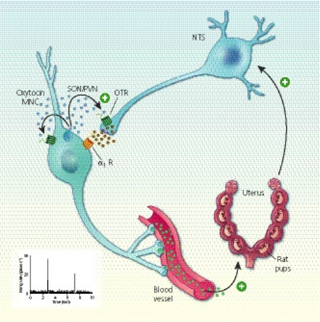
Illustration caption: "Autocrine modulation of burst firing in oxytocin magnocellular neurosecretory cells (MNCs). Cervical stretch during birth activates stretch receptors to activate A2 noradrenergic neurones in the nucleus tractus solitarius (NTS) that, in turn, activate somato‐dendritic oxytocin secretion from oxytocin MNCs. Oxytocin feeds back on oxytocin MNCs to increase excitability. Oxytocin also increases noradrenaline secretion within the supraoptic nucleus (SON) to establishe a local positive feedback loop that reinforces oxytocin MNC excitation and promotes oxytocin secretion into the circulation to trigger uterine contractions during birth. OTR, oxytocin receptor; PVN, paraventricular nucleus." Journal of Neuroendocrinology, First published: 17 April 2020, DOI: (10.1111/jne.12856)

